
Your clinic pathway
Chronic compartment syndrome diagnosis.
The clinic team arranges MRI with contrast of both calves and blood tests before your appointment. Professor Lee then reviews the findings, examines you and carries out consented compartment pressure testing at the same booked visit. Together, the findings guide your diagnosis and treatment plan.
Both calves, with contrast
The clinic’s dedicated CECS protocol.
Before and after exercise
The clinic’s panel includes CK measurements.
Part of the full picture
Findings considered with your lower-leg assessment.
Measurement with exercise
At your consultation, following assessment and consent.

See what assessment involves
Compartment syndrome diagnosis, explained.
Pressure testing with Professor Lee
Medline’s demonstration introduces the pressure-testing equipment and procedure. It includes needle testing.
Loading this video connects to YouTube. YouTube’s privacy policy applies.
Read the video overview
Professor Paul Lee introduces compartment pressure testing and demonstrates the pressure-monitoring equipment. The video discusses how symptoms during exercise can prompt further assessment. Our diagnosis page explains the current assessment pathway. English captions can be selected in the YouTube player.
A diagnosis case study
Anthony describes his experience of assessment. This educational case study includes a clinical examination; individual experiences vary.
Read Anthony’s transcript
Automatically transcribed and lightly edited for readability.
My name is Anthony Smith. I work for Anglian Water as an occupational health advisor. In my spare time I am also in the reserves as well. So I usually complete quite a bit of physical activity as part of that, which is quite different to my day role as I'm just sat down quite a lot of the time. So my level of activity increases when I go and do my reserves at the weekend.
The main symptoms I've been experiencing are quite solid calves, mainly increases during that activity after probably about a mile or so. It becomes very stiff and very solid, quite painful, restricts my running right down and as part of that also kind of once I've stopped, after probably about an hour or so, the symptoms get a bit better, but they still impact me, have an impact on sleep. I get like a pulsating calf, very uncomfortable when I'm trying to sleep. And yeah, just very stiff throughout the day, mainly as well. During driving I get the kind of sensation and pretty much all the time like a tingling sensation around the calf area.
I've had the symptoms for probably about a year now or just over a year. Initially I had some physiotherapy on the calves because they were very tight, still able to do a lot of walking. I used to do a lot of walking with weight as part of the reserves as well. But that continued following that. I kind of left it a little while before getting in contact with anyone else just in case it went off. However, during a lot of fitness tests as part of the reserves, I was getting a lot of cramping, a lot of the symptoms coming back on again.
So following that, I got in contact with our private healthcare scheme as part of Anglian Water and they referred me to MSK Doctors where I had a consultation. And then following that, I had ultrasound on my calves which didn't show anything at the time. The symptoms continued and following another period of time, I was told about this kind of procedure that I could have to identify if there was a problem.
Start with the person
Start with your symptoms and existing records
You do not need a confirmed CECS diagnosis to begin. Describe where symptoms occur, what exercise brings them on, the time or distance to onset and what changes after stopping. Include previous investigations and treatments. The clinic team coordinates your MRI and blood tests so your first appointment with Professor Lee brings those investigation results together.
You can enter the clinic pathway directly. When the ExtPress™ app becomes available, its diary and separate records for each leg will support this history: calf and thigh circumference, leg and thigh lengths, and before- and after-exercise measurements. These records support assessment; they do not establish a diagnosis.
Professor Lee’s pathway
MRI → blood tests → consultation and pressure testing → your plan.
Contrast MRI
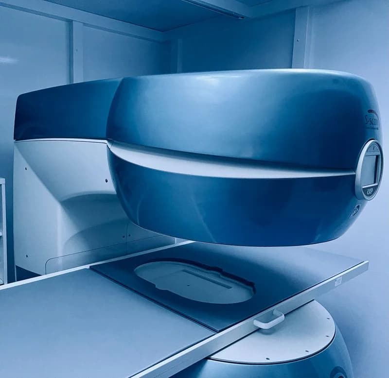
MRI with contrast of both calves
The clinic team arranges MRI with contrast of both calves using its dedicated CECS imaging protocol. If you already have an MRI, the team checks whether it covers both calves and meets the required protocol before arranging further imaging. The scan assesses the lower-leg tissues, possible abnormalities and alternative causes of symptoms. The imaging team reviews your medical history and contrast suitability before the scan.
The MRI is completed before the consultation and compartment testing, so Professor Lee has the investigation findings available to review with you.
“What are we looking for, and how would the answer change my care?”A question you can ask
Muscle response before and after exercise
Blood tests close to your MRI
The clinic team arranges blood tests close in time to the MRI and before the consultation and compartment testing. These include creatine kinase (CK) samples before and after exercise. CK is an enzyme found in muscle; the results contribute information about the muscle response to activity.
The clinic uses a specific blood panel; exercise and sample timing follow its agreed blood-testing protocol. Professor Lee brings together your symptoms, examination, MRI, blood results and pressure readings to explain the findings and recommend treatment or further investigation.
- Before exercise
A baseline CK blood sample.
- After exercise
A second sample to review the CK response.
- Findings together
CK, MRI, measurements, symptoms and pressure.
Included in your assessment
Foot scan
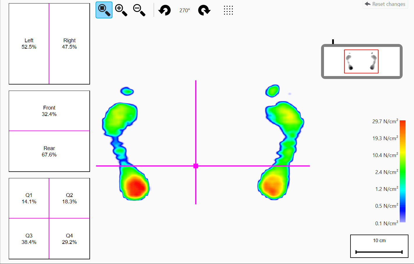
A foot scan is included in the complete assessment package. The team explains what the scan records and how the findings contribute to the assessment of your feet and lower legs.
It is one part of the wider assessment, rather than a standalone test for CECS. Professor Lee considers the findings alongside your symptoms and other investigations.
Recording a walking sequence
Reviewing balance
Reviewing a step
Exercise. Measurement. Clinical insight.
Consultation and compartment pressure testing
At your booked clinic visit, Professor Lee reviews your history, MRI, blood results and any available records, then examines you. His physical assessment includes height, the thigh-to-cleft length ratio and maximum calf circumference. Following assessment and consent, compartment pressure testing takes place during the same visit. Local anaesthetic is used for comfort.
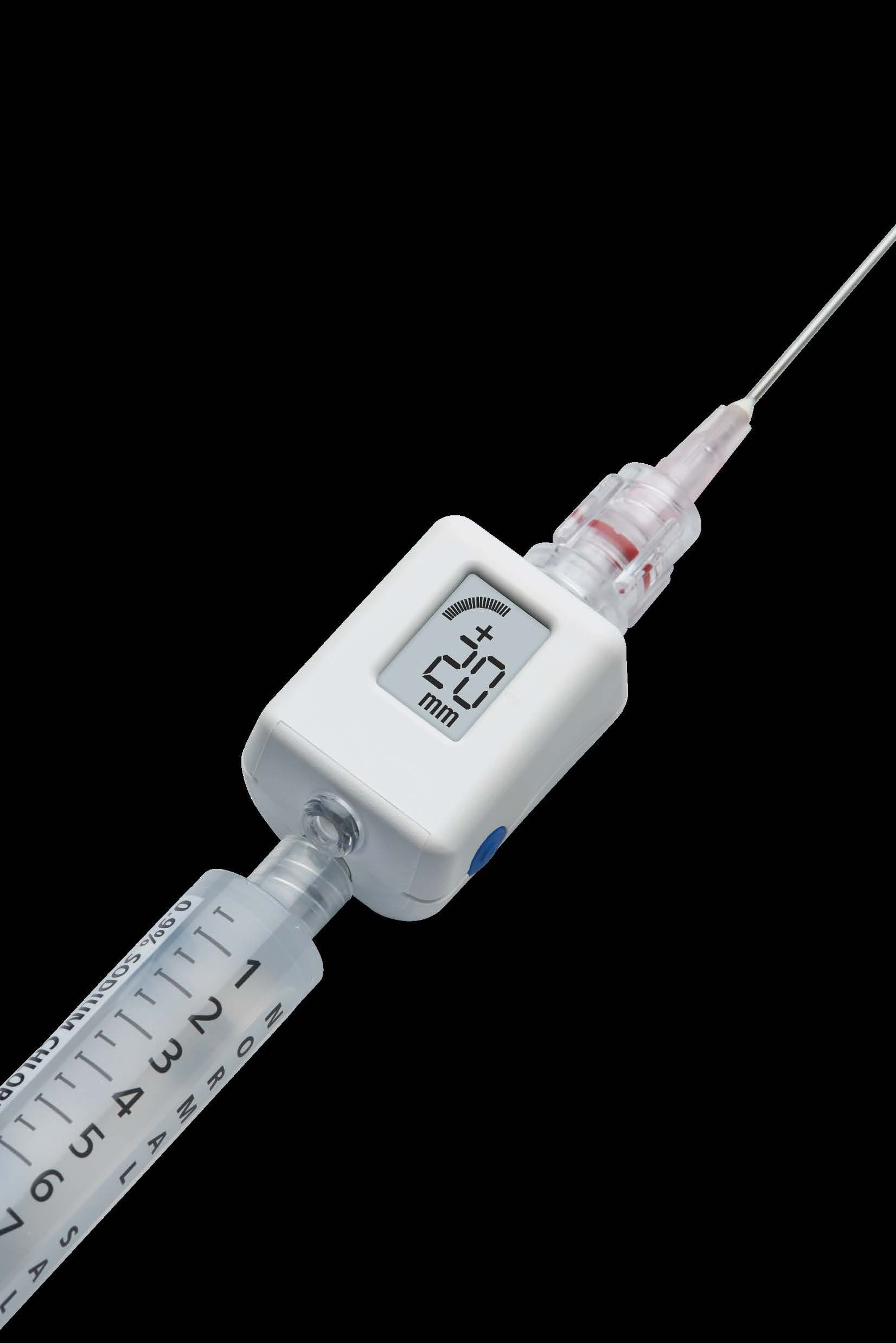
A fine needle connects to a small pressure-measuring device in each compartment being tested: anterior, lateral, superficial posterior and deep posterior. Exercise reproduces your usual symptoms. The clinic records pressure at rest, directly after exercise and five minutes after exercise.
Professor Lee interprets the pressure response alongside the other findings. The clinician explains the procedure and risks, including bleeding, infection and nerve injury. Compartment testing supports diagnosis; compartment release is a separate treatment procedure. Pressure-test protocols differ between services.
NHS: compartment syndrome · Vogels et al. (2022): clinical consensus on lower-leg CECS
Common questions
When will I see Professor Lee?
The clinic team first coordinates your MRI and blood tests. Your first appointment with Professor Lee brings those results together for review. Examination and consented compartment pressure testing take place at that same booked visit.
Is pressure testing the same as compartment release?
No. Pressure testing measures the response to exercise and supports diagnosis. Compartment release is a separate treatment that opens the fascia around affected muscles.
Do I need the ExtPress™ app before booking?
No. You can enter the clinic pathway directly, with or without the ExtPress™ app. Bring your symptom notes, previous reports and scan access details; the team explains how to provide records securely.